Picking out the best Veterinary Ultrasound Machine in 2026 isn’t just about glancing at screen sizes or shiny ads. You really need to think about what actually fits your practice—like, how many patients you see, what kind of cases you handle, and what your team is comfortable with. For example, a small, portable probe might be perfect if you’re doing mobile work with horses, but if you run a big referral hospital, you probably need something with fancy Doppler features, presets for cardiac scans, and a bunch of different transducers. It’s all about context—because, honestly, there’s no one-size-fits-all "best" out there.
Market reports from places like Grand View Research tell us the market keeps growing, thanks to more people taking care of pets, monitoring livestock, and making diagnostics easier to access. Similarly, Fortune Business Insights points out that the rising demand for vet diagnostics is fueling this growth. That’s good info, but it doesn’t tell you which machine will actually work best in your clinic. To figure that out, you'll want to dig into specs, service histories, and — ideally — actually test them out.
When you're evaluating, pay close attention to image quality—especially around the bladder wall, liver edges, and fetal structures. Try out the machine on different-sized animals. A showroom model might look amazing, but if it's being used on a restless Labrador, the real-world performance might tell a different story. For ultrasound machines for vet use, features like probe frequency, how deep the imaging goes, Doppler sensitivity, battery life, startup time, and ease of cleaning are all super important. And don’t forget support from the manufacturer—companies like Esaote, FUJIFILM Sonosite, Mindray, and Samsung all have their own strengths, whether it's portability, workflow, or image depth.
Honestly, there’s no perfect pick. Some high-end systems pack a punch but are pricey to keep running. Others are easier on the wallet but don’t offer many upgrade options. It’s smart to request a live demo, check the warranty details, and ask seasoned vets about how reliable they’ve been over the long haul. Sometimes, taking your time with the decision is the safest move—it’s better than just buying something based on slick specs alone.
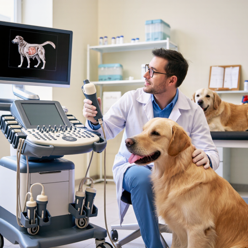
The best veterinary ultrasound machine in 2026 is defined by clinical usefulness, not impressive specifications alone. It should produce clear images across abdominal, cardiac, reproductive, and musculoskeletal examinations. A practical system needs suitable probes, reliable Doppler, and smooth depth adjustment. Operators should identify fluid, organ borders, and subtle tissue changes without fighting the controls.
Workflow matters every day. A responsive touchscreen, programmable presets, and a lightweight probe can reduce examination time. Battery performance is important during farm visits or emergency work. The machine should also tolerate frequent cleaning, accidental splashes, and continuous handling. Secure image storage and easy report transfer support communication with referral specialists. These features strengthen professional accountability.
Artificial intelligence may assist measurements, but it should not replace veterinary judgment. That assumption can fail. A useful system must display limitations clearly and preserve operator control. Training, technical support, warranty coverage, and probe availability deserve equal attention. A cheaper purchase may become expensive when repairs interrupt appointments. I would test the machine on real cases, not only display images. Ask whether a nervous dog can be scanned quietly, whether a deep-chested patient remains visible, and whether a new user can learn it safely. The best choice is the one that delivers dependable evidence under ordinary clinical pressure.
A veterinary ultrasound machine is only as useful as the images it produces at the point of care. For abdominal scans, look for clear tissue contrast, adjustable depth, and probes suited to both small patients and larger dogs. A high-frequency linear probe can reveal superficial structures, while a lower-frequency convex probe reaches deeper organs. Image quality should remain readable when a restless patient shifts or breathing blurs the screen. That matters in real clinics.
For cardiac assessment, useful features include smooth frame rates, color Doppler, and controls that make blood-flow patterns easier to evaluate. Doppler findings still need interpretation alongside the patient’s history and physical examination; a colorful display alone is not a diagnosis. Portable systems can help during bedside checks, but screen size and battery life deserve attention before purchase. A dim display in a bright treatment room is a practical problem, not a minor detail. The phrase Dog Use Ovulation Detector may appear in product searches, yet reproductive assessment and ultrasound imaging are not interchangeable tasks. Match the probe and software to the examination, and confirm that staff can save, review, and share images reliably. No machine makes every scan easy. Some cases will remain uncertain.
Choosing the best veterinary ultrasound machine in 2026 starts with the animals you examine, not a feature list. A compact Ultrasound Veterinary Machine can suit farm visits, where power outlets and quiet rooms are not guaranteed. For cattle, a durable probe and a screen that remains readable outdoors matter. Reproductive exams often need a suitable rectal probe and dependable image depth. A small-animal clinic may instead prioritize detailed abdominal imaging and easy probe changes. Fit matters.
For dogs and cats, look for clear near-field images that help distinguish small structures. A broad frequency range can support different patient sizes, but more settings do not automatically improve an examination. For equine work, consider a sturdy cart or portable unit, long battery life, and probes suited to tendon or reproductive scanning. Wildlife and mixed-practice teams may benefit from lighter equipment that is easier to carry and clean between patients. A muddy field tests design claims quickly.
Before buying, compare image quality on the species and body areas you actually scan. Ask who will provide training, service, and probe repairs, and check the total cost of ownership. Handle the controls with gloves. Are they still intuitive? I would not choose a machine from a brochure alone; a trial scan can reveal awkward menus or a screen that washes out in daylight. No single system is ideal for every species, and that trade-off deserves honest consideration.
A veterinary ultrasound system should fit the work, not just the room. Portable units are useful for farm calls, busy treatment areas, and quick checks beside a kennel. A light machine can move easily between patients, while a battery helps when outlets are scarce. The trade-off is real: a compact screen may be harder to read during a long examination. Small details matter.
Cart-based systems suit clinics that perform frequent abdominal, cardiac, or reproductive scans. Their larger displays can make it easier to compare structures and review image detail. Wheels help move the unit between rooms, though cables and narrow doorways can slow that process. Staff should test the controls, probe reach, and cleaning routine in the actual examination space before choosing.
Specialty systems are designed around particular tasks, such as cardiac imaging or high-frequency examinations of small structures. The right probe and software can be more important than a long list of features. Ask how well the system handles the patients you see most often, from a restless cat to a large-breed dog. Image quality also depends on operator training, patient positioning, and regular equipment checks. A highly capable machine will not fix an awkward workflow; even experienced teams sometimes need time to adapt.
Portable, cart-based, and specialty systems should be selected according to the transducer frequency required for each veterinary examination.
The chart shows typical transducer frequency ranges used in veterinary ultrasound. Lower frequencies generally provide greater penetration for cardiac and large-animal examinations, while higher frequencies provide better detail for superficial structures, small-animal abdominal imaging, tendons, and ophthalmic studies. Actual performance depends on probe design, patient size, depth, and system software.
In 2026, the best Veterinary Ultrasound Machine should make clinical decisions faster, not merely produce attractive images. Image quality depends on penetration, contrast, frame rate, and probe selection. A 2024 Grand View Research report estimates the veterinary ultrasound market will expand steadily through 2030, reflecting broader adoption in clinics. Yet market growth does not guarantee diagnostic value.
During a liver scan, I would examine tissue borders, small vessels, and motion artifacts at the console. Clear images matter. A responsive workflow matters more. The system should allow rapid patient registration, preset changes, measurements, and image export with minimal keystrokes. The American Institute of Ultrasound in Medicine recommends documented quality assurance and regular equipment checks. That guidance supports consistent performance, although it does not replace clinical judgment.
Mordor Intelligence’s 2024 market analysis identifies portability and point-of-care use as important growth drivers. Therefore, battery life, probe ergonomics, boot speed, and cleaning practicality deserve serious attention. A lightweight cart can still become awkward beside a nervous dog. Touchscreens may save time, but wet gloves can expose usability weaknesses. I would test the machine with real cases, not showroom clips. My early preference might change after scanning obese patients, restless cats, and deep abdominal structures. The “best” choice remains imperfect, because workflow, training, and patient cooperation alter every result.
| System Category | Typical Clinical Use | Image Quality | Workflow and Productivity | Ease of Use | Portability | Best Fit in 2026 |
|---|---|---|---|---|---|---|
| Premium cart-based system | Specialty hospitals, referral centers, cardiology, oncology, advanced abdominal and musculoskeletal examinations | Excellent detail, tissue contrast, Doppler sensitivity, and image uniformity; usually supports advanced cardiac and vascular measurements | Fast exam presets, large display, multiple probe connections, integrated measurements, DICOM connectivity, and efficient review of complex studies | Very good after training; extensive controls and advanced features require a structured learning period | Low; designed for dedicated examination rooms | The strongest choice when diagnostic image quality, repeatability, and advanced reporting are priorities |
| Advanced compact cart system | General veterinary practice, emergency care, abdominal studies, reproduction, and routine cardiac screening | High-quality 2D imaging and color Doppler for most routine small-animal examinations; generally less capable than premium systems for difficult patients and advanced cardiology | Good preset-based workflow, practical measurement packages, internal storage, and common network or DICOM export options | Good balance of guided controls, touchscreen functions, and physical buttons | Moderate; can be moved between rooms but is not ideal for field work | The best overall category for many mixed-practice clinics seeking strong capability without a full specialty-system footprint |
| Laptop-style portable system | Mobile veterinary services, farm calls, emergency response, ambulatory equine work, and clinics with limited space | Good to very good for general scanning; performance depends strongly on probe selection, battery condition, software, and operator technique | Supports digital storage, measurements, and wireless or wired export; smaller displays and fewer probe ports may slow complex examinations | Generally intuitive, although keyboard, trackpad, and screen ergonomics vary by design | High; suitable for transport and bedside scanning | A practical option when mobility and diagnostic versatility are equally important |
| Handheld probe with mobile device | Focused examinations, triage, point-of-care assessment, fluid detection, bladder checks, and field screening | Useful for focused scans; generally more limited in penetration, Doppler control, probe selection, display size, and advanced cardiac assessment | Very fast startup and easy image sharing; workflow depends on the mobile application, device compatibility, connectivity, and data-management policy | Very easy to carry and deploy; image optimization requires appropriate presets and operator training | Excellent; fits in a small case and can be used at the point of care | Best as a complementary tool for triage and rapid assessment rather than a full replacement for a diagnostic ultrasound platform |
| Dedicated equine or large-animal portable system | Reproductive examinations, tendon and ligament assessment, abdominal scanning, and ambulatory large-animal practice | Designed for deeper penetration and field conditions; image quality is optimized around specialized probes and large-animal applications | Rugged controls, long battery operation, probe options for field work, and image storage for follow-up examinations | Good in the field; physical controls are often preferable when gloves, dust, or wet conditions are involved | High, but typically heavier and more rugged than small-animal portable systems | The right choice for ambulatory large-animal work where durability, penetration, and battery life matter most |
What Is the Best Veterinary Ultrasound Machine in 2026?
A Veterinary Portable Ultrasound Machine should earn its place through measurable value, not attractive specifications. Grand View Research estimates the global veterinary ultrasound market could grow at about 6.9% annually through 2030. That growth may increase choice, but it can also confuse buyers. Compare total ownership cost, including probes, batteries, software, service visits, and staff training. A lower purchase price may become expensive after repeated repairs. Ask for documented image-quality tests, warranty terms, and average response times. Support matters.
Durability requires practical testing. Examine the handle, screen hinge, probe connectors, wheels, and battery performance in daily use. A busy clinic may scan on metal tables, farms, or uneven floors. Dust and fluid exposure are realistic concerns. Request service records from comparable users, not only laboratory claims. Future upgrades deserve equal attention. Check whether the system accepts new transducers, measurement packages, remote support, and secure data export. The 2024 Future of Veterinary Care report from the American Veterinary Medical Association emphasizes technology adoption alongside workforce efficiency, making workflow compatibility increasingly important. Still, upgrade promises can be vague. Get them in writing. I would also reserve part of the budget for operator training, because advanced features cannot correct poor scanning technique. That is an uncomfortable limitation. A technically powerful system may deliver weak clinical value when users lack practice, time, or reliable technical assistance.
The ECI-X8M veterinary ultrasound machine is designed for fast, accurate livestock diagnostics, combining a 13-inch high-definition touchscreen with multi-probe color Doppler imaging. Its probe options support examinations in pigs, cattle, sheep, and other farm animals, allowing veterinarians to assess pregnancy, reproductive status, blood flow, and cardiac function in different species. Dedicated measurement tools also assist with back-fat thickness, loin-eye muscle depth, and gestational age, supporting both breeding decisions and meat-quality management.
The scale of modern livestock production increases the need for efficient, non-invasive monitoring. According to the FAO *Statistical Yearbook 2023*, global meat production reached approximately 361 million tonnes in 2022, making reproductive efficiency and animal health important factors in supply stability. In this environment, portable ultrasound can help identify pregnancy earlier, reduce unnecessary handling, and improve breeding schedules. Color Doppler further supports evaluation of vascular activity and fetal development, while interchangeable probes provide suitable imaging depth for different animals.
The system is particularly valuable for farms, veterinary suppliers, and agricultural service providers seeking consistent field performance. Its touchscreen workflow enables clear image review and convenient measurements during routine herd examinations, while multi-function imaging supports reproduction management, growth assessment, and cardiac screening in one platform.
Clinical usefulness matters more than impressive specifications. It should show clear organs, fluid, vessels, and tissue changes. Reliable probes and simple controls are essential.
Choose equipment for the animals and body areas you scan most. Dogs and cats need detailed abdominal images. Horses may need tendon and reproductive probes. Farm visits require portability and battery power.
A lightweight system, strong battery, readable outdoor display, and durable probe are valuable. Power outlets may be unavailable. Mud tests every design claim.
Both are critical. Check penetration, contrast, frame rate, and near-field detail. A suitable probe can reveal small structures more clearly. More settings do not guarantee better results.
Fast booting, programmable presets, quick measurements, and easy image export help reduce delays. Controls should remain clear with gloves. Wet fingers can expose weaknesses.
No. It may assist measurements, but the veterinarian must interpret the evidence. Its limits should be visible. That assumption can fail.
Request real scans with nervous dogs, restless cats, obese patients, and deep-chested animals. Check image visibility in daylight. Handle the controls yourself, not only the brochure.
Ask about training, repairs, warranty coverage, cleaning tolerance, and probe availability. A cheaper unit may become costly during repeated repairs. My preference might change after service delays.
No single system fits every species, workload, and budget. A portable unit may suit field work, while a clinic may need deeper imaging. The trade-off deserves honest consideration.
The best veterinary ultrasound machine in 2026 is defined by its ability to deliver clear, reliable images while supporting efficient diagnosis across a wide range of clinical situations. Important features include high-resolution imaging, Doppler capabilities, flexible probes, adjustable settings, and software that helps veterinarians assess soft tissue, abdominal organs, pregnancy, cardiac function, and emergency cases. The right Ultrasound Machine Veterinary Use should also match the needs of the animal species being examined, from small companion animals to large animals and specialized patients.
Portability, cart-based mobility, and specialty configurations each offer practical advantages. A strong system should combine image quality with an intuitive workflow, fast operation, ergonomic design, and easy data management. Buyers should also evaluate total cost, durability, maintenance requirements, technical support, training, and opportunities for future upgrades. Ultimately, the best choice is not simply the most advanced or expensive machine, but the one that provides dependable performance, supports daily clinical efficiency, and can adapt to changing veterinary practice needs.