In the world of veterinary medicine, which keeps changing all the time, the Vet Ultrasound Scannerreally stands out as such a vital tool for diagnosis. Dr. Emily Carter, who’s pretty well-known at Animal Health Innovations, often points out that "the Vet Ultrasound Scanner gives an incredible look into what’s going on inside a pet’s body." Basically, it helps vets see beyond what's just on the surface — kind of like a peek inside without chopping into anything.
This device uses sound waves to generate images of organs and tissues inside an animal—think of it like an echo that creates pictures. What’s cool? It’s non-invasive, so it’s great for regular check-ups or when something urgent comes up. Plus, it can spot issues that X-rays or physical exams might miss. For example, vets can check out the heart, kidneys, and even detect tumors in real-time, right there in the exam room.
That said, there’s still some room for improvement — especially when it comes to clarity and accuracy across different sizes of animals. And let’s be honest — some pet owners find the whole process a little intimidating at first, which really highlights how important it is to educate folks more about how this tech works. Still, without a doubt, it’s a key part of vet work today. Learning how this scanner functions can actually help pet parents feel more confident and better equipped to make decisions about their furry friends’ health.
What is a Veterinary Ultrasound Scanner?
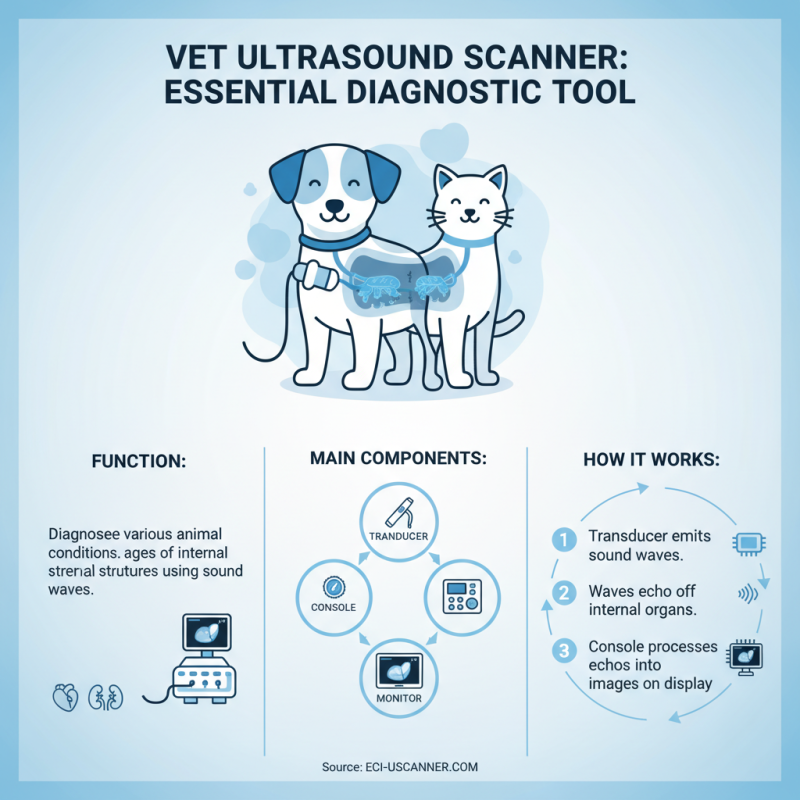
Veterinary Ultrasound Scanners are essential tools in modern animal healthcare. These devices utilize sound waves to create images of internal structures. Unlike traditional X-rays, ultrasounds do not use harmful radiation. This makes them safer for pets and animals. The scans are useful for diagnosing conditions in various species, from dogs to cats, and even livestock.
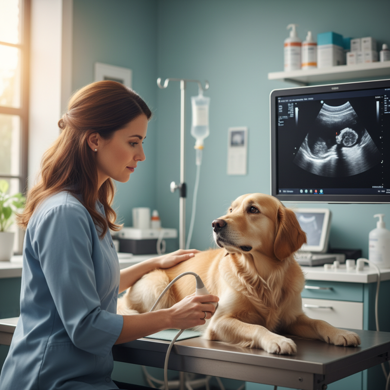
Using a vet ultrasound scanner involves a few steps. A gel is applied to the animal's skin to enhance sound wave transmission. The probe, or transducer, is then moved over the area of interest. The images are displayed in real-time, allowing veterinarians to see organs and tissues in detail. This process can reveal issues such as tumors, fluid abnormalities, or pregnancies. However, it requires skill and practice to interpret the results accurately.
Despite the advancements, challenges remain. Some animals may struggle with anxiety during the procedure. Additionally, certain conditions can be difficult to visualize. The quality of the images depends on various factors, including the animal's size and the technician's experience. Each scan is a learning experience for both the veterinarian and the pet owner. With each case, there’s room for reflection and improvement in techniques.
What is a Vet Ultrasound Scanner and How Does It Work?
| Feature | Description | Benefits |
| Image Clarity | Provides high-resolution images of soft tissue structures. | Facilitates accurate diagnosis of internal conditions. |
| Non-Invasive | The procedure is safe and does not require incisions. | Reduces risk associated with surgical diagnostics. |
| Real-Time Imaging | Enables live movement observation of internal organs. | Allows for dynamic assessment of organ function. |
| Portable Options | Some models are lightweight and easy to transport. | Useful for fieldwork and emergency situations. |
| User-Friendly Interface | Designed with intuitive controls and display options. | Facilitates ease of use for veterinarians. |
Principles of Ultrasound Imaging in Veterinary Medicine
Ultrasound imaging is essential in veterinary medicine. It uses sound waves to create images of a pet's internal organs. A vet ultrasound scanner sends these waves into the body. When they bounce back, a computer processes the data. This process creates real-time images. Vets can monitor vital organs such as the heart, liver, and kidneys.
Understanding the principles behind ultrasound is important. The sound waves are at frequencies higher than humans can hear. They penetrate tissues and reflect differently based on density. For instance, the heart's chambers look different compared to muscle or fat. Not all tissues reflect sound equally. Sometimes, this leads to unclear images. Vets must adjust settings based on the animal's size and condition.
While ultrasound is non-invasive, it's not foolproof. There are limitations in visualizing certain conditions. Gas-filled organs can obscure images. It's vital for vets to have training to interpret complex results. Continuous learning is key. Each ultrasound offers new challenges and insights.
Common Uses of Veterinary Ultrasound in Diagnosing Animal Health
Veterinary ultrasound is a crucial tool for assessing animal health. It utilizes sound waves to create images of internal organs. This non-invasive technique helps veterinarians diagnose various conditions without performing surgery.
One common use of ultrasound is in pregnancy checks for animals. Veterinarians can visualize developing fetuses early on. This helps track the health of both the mother and her young. Another major area is in evaluating soft tissue injuries. Muscles and tendons can be scanned to detect tears or abnormalities. This imaging gives clear insights, unlike other techniques that might miss subtle issues.
Ultrasound is also essential for examining heart functions. By observing blood flow and cardiac structure, vets can identify problems. Some animals may respond well to treatments, while others may require more invasive procedures.
Limitations exist, though. Images can be affected by patient movement or excessive fat. This requires the vet to constantly adapt their approach, seeking clearer views.
Key Components of a Vet Ultrasound Scanner Explained
Vet ultrasound scanners are essential tools for veterinarians. These machines help in diagnosing a range of conditions in animals. The main components of a Vet Ultrasound Machine include a transducer, a console, and a display monitor. The transducer emits sound waves and receives the echoes. This process creates images of the internal structures of the animal.
When using a vet ultrasound scanner, the images appear almost immediately. This allows for quick diagnosis. The console is the brain of the machine, processing the signals from the transducer. It displays the images in real-time. The display monitor shows these images, making it easier for veterinarians to analyze the condition of the patient.
Tips: Ensure the animal is calm before the procedure. A relaxed pet will yield better images. Keep the ultrasound gel warm; cold gel can be uncomfortable. Familiarize yourself with the settings of the Vet Ultrasound Machine for optimal use. Each animal may require a different approach, and sometimes the images may not be perfect. Don’t hesitate to repeat a scan if necessary; clarity is vital for accurate diagnosis.
How to Choose the Right Ultrasound Scanner for Veterinary Practice
Choosing the right ultrasound scanner for veterinary practice can be daunting. The market offers many options, each with varying features and capabilities. According to the American Veterinary Medical Association (AVMA), the demand for ultrasound in veterinary medicine has increased by 30% over the last five years, emphasizing its importance in diagnostics.
When selecting an ultrasound scanner, consider factors like portability, image quality, and ease of use. A Portable Vet Ultrasound device can significantly enhance mobility and accessibility, especially for emergency cases or large animal practices. Experts recommend models that support both 2D and Doppler imaging to expand diagnostic capabilities. However, not all portable models deliver the same image quality, leading to potential misdiagnoses.
It's also crucial to evaluate training resources and customer support when making a purchase. A scanner might be impressive, but inadequate support can hinder the learning curve. Difficulty in navigating user manuals or software can create frustration. Many practitioners report that finding a balance between advanced features and user-friendly interfaces is a common challenge. Selecting an ultrasound scanner is more than just a financial investment; it impacts your ability to provide quality care.
Industry Trends: Advancements in Veterinary Ultrasound Technology
Advancements in veterinary ultrasound technology are transforming animal healthcare. Today's equipment is more portable and user-friendly. The Portable Vet Ultrasound Machine, for example, allows veterinarians to perform scans in various locations. This mobility enhances patient care significantly.
The technology has evolved rapidly, yet many vets still face challenges. Not every clinic can afford the latest devices. Training staff is another hurdle. Mastering this technology takes time and practice. An effective ultrasound requires not just advanced machines, but skilled operators too.
Resolution and depth of imaging have improved drastically. However, even with advanced tools, interpretation can sometimes be unclear. Clinicians may need to rely on their experience more than the machine's capabilities. It highlights the necessity for ongoing education in this field. Ultimately, while the advancements are promising, they also require careful implementation and constant learning.
Advancements in Veterinary Ultrasound Technology
This bar chart represents the advancements in various veterinary ultrasound technologies over the years, highlighting the increase in applications and capabilities.
Impact of Ultrasound Diagnostics on Animal Care and Treatment Outcomes
Veterinary ultrasound has transformed animal care dramatically. A vet ultrasound scanner provides real-time images of internal organs. This non-invasive technique aids in diagnosing various conditions in pets and livestock alike. Using sound waves, the scanner creates detailed images that help veterinarians assess health issues. It's a clear window into the animal's body without needing surgery.
The impact of ultrasound diagnostics on treatment outcomes is significant. Quick and accurate diagnoses lead to timely interventions. Conditions that once required exploratory surgery can now be managed more conservatively. For instance, a simple ultrasound can reveal tumors or fluid buildup. However, the technology is not foolproof. Misinterpretation of images can happen, leading to unnecessary treatments. Veterinarians must remain vigilant in their analysis.
Moreover, having access to a Vet Diagnostic Ultrasound Machine doesn’t guarantee better outcomes. It’s crucial for vets to combine their expertise with the technology. Continuous education and experience play vital roles in effective diagnostics. While this tool enhances care, the human element is irreplaceable. Balancing technology with intuition is key.
Exploring the Benefits and Features of the CW-3C Waterproof Wireless Convex Ultrasound Probe Scanner
The CW-3C Waterproof Wireless Convex Ultrasound Probe Scanner embodies cutting-edge digital imaging technology, providing users with crystal-clear imaging that enhances diagnostic accuracy. This advanced tool leverages modern technological advancements to ensure high-resolution images that are essential for effective medical evaluations. The clarity of the imaging is particularly beneficial in clinical settings, where precision can significantly influence patient outcomes.
Designed with portability in mind, the CW-3C features a compact and lightweight structure that makes it easy to carry and use in various environments. The wireless functionality eliminates the need for cumbersome probe cables, allowing for greater mobility and flexibility during examinations. This enables healthcare professionals to work freely without being tethered to a power source, enhancing the efficiency of their workflow. Overall, the combination of high-cost effectiveness and state-of-the-art features makes the CW-3C a valuable asset in any medical toolkit, ensuring that practitioners can deliver top-notch care with ease.
FAQS
: The guide aims to provide practical advice on effective communication techniques.
Focus on the speaker, avoid distractions, and ask clarifying questions.
It conveys emotions and attitudes, often more than words can express.
Practice regularly, use positive self-talk, and prepare your points in advance.
Stay calm, listen actively, and seek common ground to resolve issues.
Clear messages, mutual understanding, and a positive atmosphere indicate success.
Yes, emotions can influence how messages are received and interpreted.
Use simple language, be specific, and organize your thoughts logically.
Feedback helps clarify understanding and can improve future interactions.
Record conversations, seek input from others, and evaluate your successes and challenges.
Conclusion
A Vet Ultrasound Scanner is a vital tool in veterinary medicine, utilizing sound wave technology to create images of an animal's internal structures. The principles of ultrasound imaging rely on the transmission of high-frequency sound waves that reflect off tissues, providing clear visuals for diagnosing various health conditions. Its common applications include assessing organ health, detecting tumors, and monitoring pregnancies in animals.
When selecting an appropriate Vet Ultrasound Scanner for veterinary practice, it is essential to consider key components such as image quality, portability, and user-friendly interfaces. The advancements in veterinary ultrasound technology continue to enhance diagnostic capabilities, significantly improving animal care and treatment outcomes. By integrating ultrasound diagnostics, veterinarians can make informed decisions, leading to more effective treatments and better overall health for their patients.

 Bovine
 Equine
 Swine
 Ovine
 Pet