Bovine
 Equine
 Swine
 Ovine
 Pet





How Many Types of Ultrasound Machines Are There?
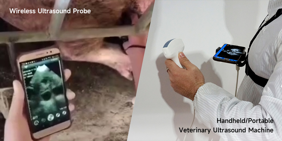
Based on the physical design and mobility of the main unit, veterinary ultrasound machine can be primarily categorized into four types: Wireless Ultrasound Probe, handheld/portable vet ultrasound, laptop-style/notebook animal ultrasound, and cart-based/console veterinary ultrasound.

Design: A standalone probe without an integrated screen. It typically resembles a large mouse or an oval-shaped device, connecting to smart terminals (like tablets or smartphones) via wireless technology.
Size: Extremely compact and lightweight, often weighing only a few hundred grams, making it easily pocketable. Hence, wireless ultrasound probe is also referred to as a palm or pocket ultrasound.
Application: Due to its small size and portability, it is ideal for mobile practices and rapid on-site screening in settings like farms. wireless ultrasound probe is primarily used for early pregnancy detection in small and medium-sized animals (e.g., dogs, cats, sheep, pigs) and examination of superficial tissues.
Handheld/Portable Veterinary Ultrasound Machine
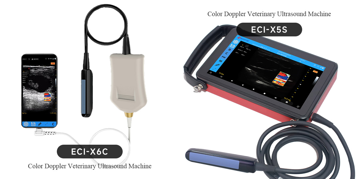
Design: The key feature is a main unit (integrating the processor and display) designed to be held in one hand. The main unit is usually square or rectangular, connected to an external probe via a cable. The operator holds the main unit in one hand and manipulates the probe with the other.
Size: The main unit display typically ranges from about 5 to 13 inches. The entire setup is lightweight, usually weighing between 1 to 2 kg including the probe.
Application: Portable veterinary ultrasound machine offers an optimal balance between portability and functionality. It is suitable for outdoor visits, pen-side examinations on farms, and emergency or general consultations in veterinary clinics. Portable Animal Ultrasound Machine adequately meets needs such as pregnancy diagnosis, back-fat measurement, and examination of some abdominal organs.
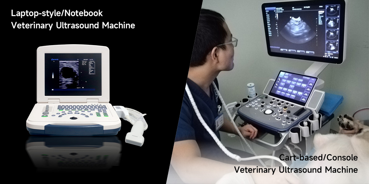
Laptop-style/Notebook Veterinary Ultrasound Machine
Design: Resembles a laptop computer, featuring a clamshell design with a display unit and a separate keyboard. The probe connects to the side or rear of the main unit via a cable.
Size: Larger in volume, typically weighing between 3 to 6 kilograms. It often comes with a carry handle or dedicated backpack for easier transportation.
Application: Suitable for fixed clinics, medium-sized farms, or veterinary teams that require full functionality while moving frequently between locations. Laptop-style/Notebook veterinary ultrasound machine provides a larger screen and more powerful processing capability, making it suitable for more detailed examinations of the abdomen, heart, tendons, etc.
Cart-based/Console Veterinary Ultrasound Machine
Design: A large, integrated workstation typically consisting of a high-performance main unit, a large professional display (≥15 inches), and multiple probe ports, all mounted on a multi-functional trolley with wheels.
Size: This is the largest and heaviest category. Mobility relies on the trolley, making it best suited for movement within a fixed space like a clinic.
Application: Primarily used in large animal hospitals, veterinary teaching and research institutions, and high-performance equine or dairy facilities. Cart-based/Console veterinary ultrasound machine supports comprehensive advanced applications, including color Doppler, spectral Doppler, and high-frequency ultrasound, essential for specialized fields like cardiology, ophthalmology, deep tendon diagnostics, and research.
Note: As ultrasound technology continues to be upgraded, mobile palm and portable veterinary ultrasound machines are also equipped with high-end color ultrasound and Doppler capabilities.
For further details, please contact us.
Classification by Imaging Technology
Veterinary ultrasound machine can also be classified based on its core imaging principles and technology into A-mode ultrasound, B-mode (Black & White) ultrasound, and Color Doppler ultrasound.
Imaging Principle: Amplitude Modulation. This is an early technology that displays information as a one-dimensional waveform graph, used for measuring distance and organ size.
Core Application: Primarily used for precise measurement. In veterinary medicine, its classic application is measuring back-fat thickness in livestock (e.g., pigs) to evaluate carcass quality and lean meat percentage. It can also be used for rough localization of fluid-filled lesions (e.g., cysts).
Current Status: A-mode veterinary ultrasound functionality is limited. It has largely been integrated into or replaced by the more intuitive B-mode veterinary ultrasound and is now primarily used as a specialized tool for specific production performance measurements.
B-mode (Black & White) Veterinary Ultrasound Machine
Imaging Principle
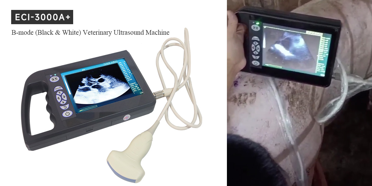
B-mode (Brightness-mode): This is the dominant mainstream mode. Multiple ultrasound beams create a cross-sectional scan. Echo signal strength is converted into points of varying brightness, forming a two-dimensional grayscale anatomical image on the screen. This clearly displays organ shape, boundaries, internal structure, and lesions.
M-mode (Motion-mode): This mode displays motion over time. A sampling line is placed on the B-mode image, and the movement of structures along that line is plotted against time (vertical axis: depth, horizontal axis: time).
Function & Characteristics: B-mode ultrasound is the cornerstone tool in veterinary diagnostics for visualizing the morphological structure of nearly all organs.
Main Applications Include
Reproductive Management: Pregnancy diagnosis, litter size determination, fetal development monitoring, and assessment of uterine/ovarian disorders.
Abdominal Examination: Evaluation of the size, shape, and structure of organs like the liver, gallbladder, spleen, kidneys, and bladder for abnormalities.
Other Uses: Examination of tendons, assessment of superficial masses, and detection of pleural or abdominal effusion.
Functional Assessment: M-mode is primarily used in cardiac examination to accurately measure heart chamber dimensions, ventricular wall thickness, and motion amplitude, making it a key tool for evaluating cardiac function.
Color Doppler Veterinary Ultrasound Machine
Imaging Principle: Based on the Doppler Effect, specifically used to detect and display blood flow information.
Main technologies include
Color Doppler Flow Imaging (CDFI): Color is superimposed on the real-time B-mode grayscale image to indicate blood flow direction (typically red towards the probe, blue away) and velocity (indicated by color brightness), providing a visual map of vascularity and flow status.
Spectral Doppler: This mode samples flow at a specific point and displays the change in blood flow velocity over time as a waveform graph, allowing precise quantitative measurements like flow velocity and resistance indices.
Function & Characteristics
Hemodynamic Assessment: Color Doppler veterinary ultrasound machine provides a powerful functional complement to the morphological diagnosis of B-mode ultrasound.
Main Applications
Cardiac Diagnosis: Essential for assessing valvular regurgitation/stenosis, congenital heart defects, cardiomyopathy, etc., making it indispensable in veterinary cardiology.
Vascular Examination: Diagnosis of vascular thrombi, stenosis, aneurysms, etc.
Tumor Differentiation: Assists in distinguishing between benign and malignant tumors by observing the patterns and richness of blood flow within and around a mass.
Organ Perfusion Assessment: Evaluates blood flow to organs like the kidneys and liver.